We have applied some changes to ARIA and are monitoring stability, but you can continue to work as normal. We appreciate your understanding.
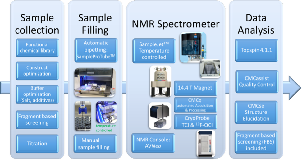
BMRZ has significantly expanded its capacity in fragment- and drug-screening for both RNAs and proteins. Two dedicated spectrometers (600 MHz) are optimised for screening using 1.7 mm, 3.0 mm, and 5.0 mm probes, including specialised ¹⁹F and low-gamma detection probes.
BMRZ has significantly expanded its capacity in fragment- and drug-screening for both RNAs and proteins. Two dedicated spectrometers (600 MHz) are optimized for screening using 1.7 mm, 3.0 mm, and 5.0 mm probes, including specialized ¹⁹F and low-gamma detection probes.
The BMRZ has significantly expanded its capacity for fragment- and drug-screening of both RNA and protein targets. Two dedicated 600 MHz NMR spectrometers are optimized for screening applications and equipped with 1.7 mm, 3.0 mm, and 5.0 mm probes, including specialized 19F and low-γ detection probes.
Researchers can perform screening campaigns using either:
These resources, together with the automated NMR infrastructure, enable high-throughput and reproducible fragment-screening workflows for a wide range of biomolecular targets.
Sample Preparation Guidelines
For each screening experiment, prepare two types of NMR samples:
NMR tube options and sample volumes:
For sample setup, use standard Bruker NMR tubes compatible with HT sample changers.
Each NMR tube’s barcode ID is automatically linked to the acquired dataset, ensuring a fully traceable, ID-based workflow. The robotic temperature-control system maintains constant conditions during sample preparation and measurement.
Further Information and Support
For detailed methodology and step-by-step setup, users are strongly encouraged to consult the Facility’s publication in the Journal of Visualized Experiments (JoVE):
“NMR-Based Fragment Screening in a Minimum Sample but Maximum Automation Mode” DOI: 10.3791/62262
Before planning or conducting any screening experiment, please discuss your project with the Facility’s scientific experts to ensure optimized, target-specific screening conditions: Christian Richter and Sridhar Sreeramulu (Early consultation with the team helps develop a user-tailored screening strategy, ensuring efficient use of available instrumentation, samples, and screening resources.)
Availability: yes
Physical access: yes
Remote access: yes
Average time of a visit 1-2d